Advanced Technology. Earlier Detection. More Personalized Care.
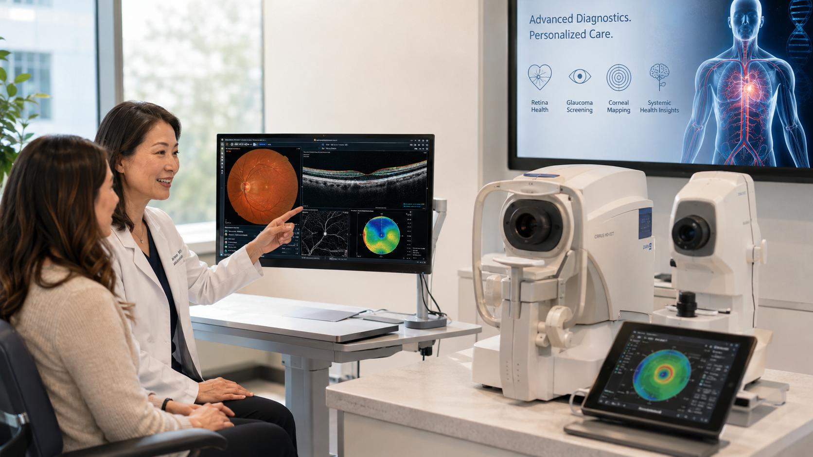
At High Point University Eye Health, advanced technology is an important part of how we protect your vision and care for your overall eye health. Our clinic is equipped with sophisticated diagnostic and imaging systems that allow our doctors to see structures of the eye in remarkable detail, often identifying subtle changes before they cause noticeable symptoms.
These technologies help us detect eye disease earlier, establish an accurate baseline of your eye health, monitor changes over time, and develop individualized treatment plans based on what we see.
Your eyes can also provide important clues about your overall health. Changes in the retina, retinal blood vessels, optic nerve, and other structures may be associated with systemic conditions such as diabetes, high blood pressure, vascular disease, and certain neurologic conditions. When concerning findings are identified, our doctors can recommend appropriate follow-up or communication with your other health care providers.
Our care is faculty-led, with student clinicians participating under the supervision of our doctors as the School of Optometry’s clinical education program develops.
Request an AppointmentOur Advanced Diagnostic Technology
Optical Coherence Tomography (OCT)
OCT gives our doctors highly detailed, cross-sectional images of the retina, macula, and optic nerve, similar to being able to view the individual layers of these structures rather than simply looking at their surface.
OCT can help identify and monitor conditions including glaucoma, macular degeneration, diabetic eye disease, macular swelling, and other retinal and optic nerve disorders. Because OCT measurements can be compared over time, even subtle changes may be detected and monitored before significant vision loss occurs.
OCT Angiography (OCT-A)
OCT-A takes retinal imaging a step further by providing detailed images of the tiny blood vessels within the retina and surrounding tissues without the need for injected dye.
By evaluating blood flow and changes within the retinal circulation, OCT-A can help our doctors identify abnormal or reduced blood flow associated with conditions such as diabetic retinopathy, macular degeneration, retinal vascular disease, and other disorders affecting the eye’s circulation.
Because the health of retinal blood vessels can sometimes reflect broader vascular health, these images may also provide clues that warrant additional medical evaluation.
Ultra-Widefield Retinal Imaging
Traditional retinal examination provides an important view of the back of the eye. Ultra-widefield imaging allows us to capture a much broader view of the retina, including areas far into the peripheral retina that can otherwise be difficult to document.
This technology can help identify changes associated with diabetic retinopathy, retinal tears or detachments, vascular abnormalities, inflammation, and other retinal conditions.
The images also create a valuable baseline that allows our doctors to compare your retina from one visit to the next and recognize changes that may otherwise be difficult to detect.
A-Scan and B-Scan Ocular Ultrasound
Ultrasound allows our doctors to evaluate the eye using sound waves, providing important information when structures cannot be adequately seen through traditional examination or imaging.
A-scan ultrasonography provides precise measurements of the eye and can help evaluate the size and position of ocular structures.
B-scan ultrasonography creates a two-dimensional image of structures inside and behind the eye. It can be especially valuable when the view of the retina is limited by conditions such as a dense cataract, bleeding inside the eye, or other media opacity.
B-scan imaging can assist in evaluating conditions such as retinal detachment, vitreous abnormalities, masses, and other structural changes within the eye.
Corneal Topography
Every cornea has its own unique shape. Corneal topography creates a detailed map of the curvature and contour of the front surface of your eye.
This information can help our doctors detect conditions such as keratoconus and irregular astigmatism, evaluate changes in corneal shape, and provide more precise care for patients who need specialty contact lenses.
Detailed corneal measurements can also help us better understand why a patient may experience blurred or distorted vision even when a traditional prescription does not tell the whole story.
Visual Field Analysis
Your vision includes much more than what you see directly in front of you. Visual field testing measures your central and peripheral vision and can identify areas where sensitivity has been reduced.
Visual field testing is particularly important in the diagnosis and monitoring of glaucoma and optic nerve disease. Certain patterns of visual field loss may also suggest neurological conditions that require additional evaluation.
By repeating testing over time, our doctors can determine whether vision remains stable or whether a condition is progressing.
Advanced Dry Eye Evaluation and Treatment
Dry eye disease is often more complex than simply not producing enough tears. In many patients, the problem involves the quality of the tear film or the function of the oil-producing glands within the eyelids.
Our technology allows us to evaluate the underlying cause of your symptoms and develop a treatment plan designed specifically for your eyes.
Meibography
Meibography provides detailed images of the meibomian glands, the tiny glands within the eyelids responsible for producing the oils that prevent tears from evaporating too quickly.
This painless, non-invasive technology allows our doctors to evaluate gland structure and identify signs of meibomian gland dysfunction (MGD), one of the most common contributors to dry eye disease.
Identifying gland changes early can help us recommend treatment designed to preserve gland function, improve the stability of the tear film, and relieve symptoms such as burning, irritation, redness, and fluctuating vision.
OptiLift®
OptiLift® is a non-invasive treatment designed to stimulate the muscles and tissues surrounding the eyes and eyelids.
For appropriately selected patients, improving eyelid function may support healthier blinking and more effective distribution of the tear film across the surface of the eye. OptiLift may be incorporated into an individualized treatment plan for patients experiencing symptoms related to dry eye or meibomian gland dysfunction.
OptiLight®
OptiLight® uses precisely controlled intense pulsed light technology as part of the treatment of dry eye disease associated with meibomian gland dysfunction.
Treatment targets inflammation around the eyelids and can help improve meibomian gland function and tear-film stability. For appropriate patients, a series of treatments may help reduce symptoms such as burning, redness, irritation, dryness, and fluctuating vision.
Seeing More Means Caring for More
Advanced technology does not replace a comprehensive eye examination—it enhances it.
By combining detailed imaging, sophisticated diagnostic testing, and the clinical expertise of our doctors, High Point University Eye Health can evaluate not only how well you see today, but also the health of the structures responsible for maintaining your vision for years to come.
Our goal is simple: detect problems earlier, understand them more completely, and provide the personalized care needed to protect your vision and support your overall health.
Request an Appointment